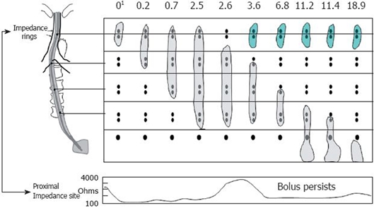
Figure 1.

Example of retrograde escape and proximal stasis on simultaneous multichannel intraluminal impedance and fluoroscopy. The fluoroscopic bolus is represented by the gray bolus at each time interval during the swallow and the green bolus during retrograde escape. The bottom panel is a single impedance tracing at the proximal location. The bolus is present at the proximal impedance site until 2.6 s where the bolus tails moves distal to the two rings at that level and the impedance tracing returns to baseline. At 3.6 s, the bolus (green) reenters the recording site and once again the impedance drops consistent with bolus retention. Modified from Imam et al[7]. 1Time in seconds.
