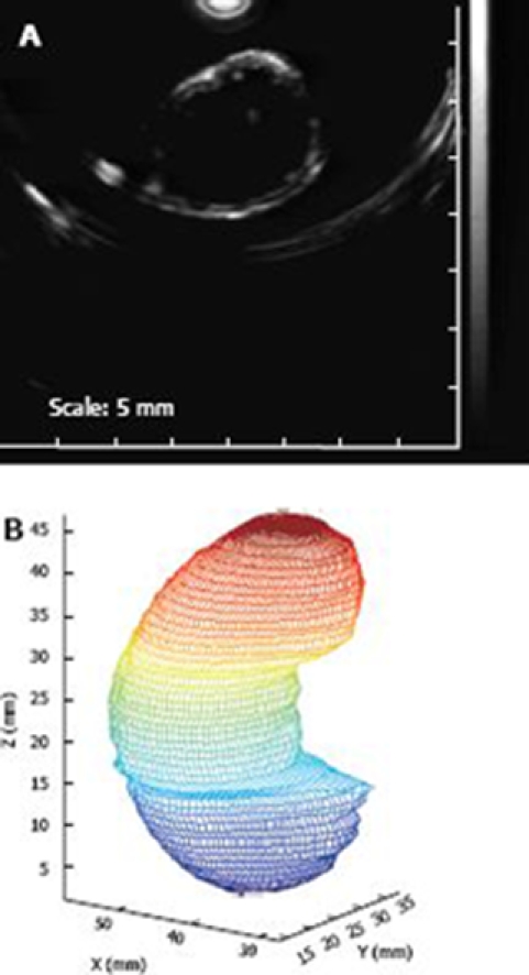
Figure 4.

An example of the anatomically based in vitro rat stomach model generated from ultrasonic scanning. A: A representative CT scanning of a cross sectional slice of an in vitro rat stomach; B: The reconstructed gastric model on the basis of the CT scanning on the in vitro rat stomach. The distance between cross sectional slices was 1 mm, the colour change from blue to red means the increase of the stomach length in z direction.
