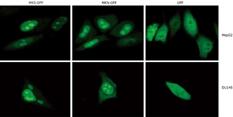
Figure 1.

Subcellular localization analysis of MKS-GFP and MKN-GFP fusion proteins in carcinoma cell lines. MKS-GFP and MKN-GFP fusion proteins were localized to nuclei and accumulated in nucleoli, but rarely localized to cytoplasm. The high light “spots” in the center of nuclei are nucleoli. HepG2 cells (upper panel) and DU145 cells (bottom panel) are transfected with pEGFP-N2, and diffuse freely in the whole cell except in nucleoli.
