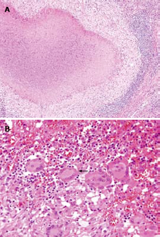
Figure 4.

Histological image of resected specimen. A: Photograph showing the epithelioid granuloma with central caseous necrosis (HE, original magnification, × 100); B: multinucleated Langhans’ giant cell with surrounding lymphocytes (arrow) (HE, original magnification, × 400).
