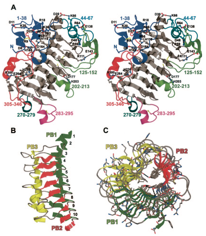
FIG. 1. Structure of Jun a 1.
A, stereo view showing the loops and the salt bridges (ball and sticks) we identified. The color of the numbers and the carbon atoms of the salt bridges refer to the loop of the same color. The three disulfides and free sulfhydryl are shown as sticks with black colored carbons. Single letter amino acid abbreviations are used with position numbers. B, parallel β-helical structure of Jun a 1 with loops removed for clarity. Secondary structures are colored as follows: green, parallel β-helical sheet PB1; red, PB2; yellow, PB3. Numbers refer to the PB sheet strand from the N (top) to C (bottom) termini. C, previous figure rotated 90° about the horizontal axis. The view is from the C terminus toward the N terminus of the β-helical core.

