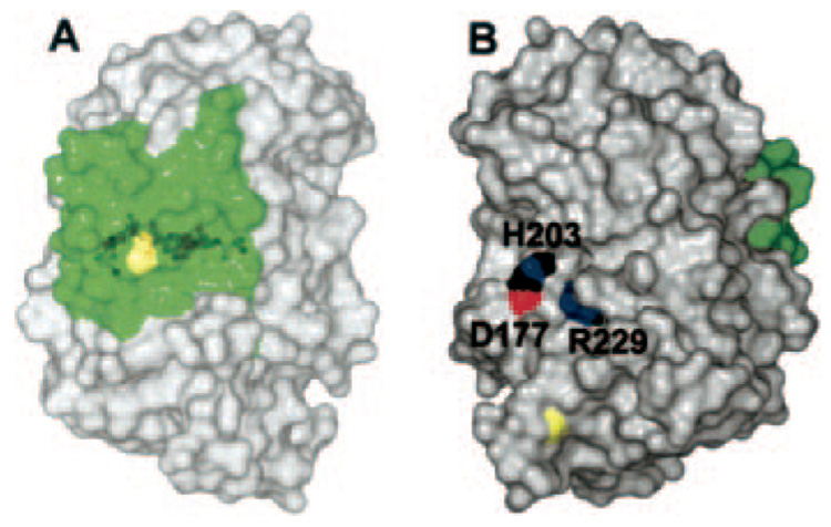
FIG. 5. The surface of Jun a 1 showing the locations of the RXPXXR and aWiDH sites.
A, the aWiDH residues covered by residues 1–30 (green). Yellow depicts the disulfide bond between Cys7 and Cys27. B, view of panel A rotated 90° about the vertical axis showing the potential substrate binding groove around the active site. His203 (H203) and Arg229 (R229) are shown in Corey-Pauling-Koltun colors. Yellow depicts the disulfide bond between Cys285 and Cys291. The aWiDH site is the green area on the right. D177, Asp177.

