Fig. 4.
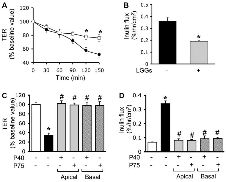
Probiotic factors at both apical and basal surfaces prevent H2O2-induced paracellular permeability. A and B: Caco-2 cell monolayers were incubated with ([□] and solid bar) or without (● and shaded bar) LGG-s for 60 min and washed twice to remove traces of probiotics prior to administration of H2O2. TER (A) was measured at varying times, and inulin flux (B) was measured at 2 h after H2O2 administration. Values are means ± SE (n = 6; values from 3 independent experiments). *Significantly (P < 0.05) different from corresponding values for cell monolayers without LGG-s treatment. C and D: Caco-2 cell monolayers were incubated with p40 or p75 (1.0 μg/ml) on the apical or basal surfaces for 30 min prior to administration of H2O2. TER (C) and inulin flux (D) were measured at 2 h after H2O2 administration. Values are means ± SE (n = 6). *Significantly (P < 0.05) different from corresponding control values; #different from corresponding values for H2O2-treated cells without p40 or p75.

