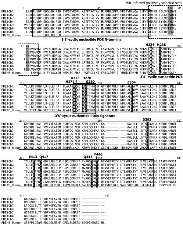
Figure 3.
Protein sequence alignment of multiple stickleback PDE1Cb and human PDE4B. The PDE tertiary structure and active enzyme sites (black shading) are reported for the human PDE4B (site numbers of active sites are according to ref. [28]). The signature domains of PDE protein are designated by solid boxes, and the sequence region that contains the ML-inferred positively-selected sites is designated by a dashed box. The stars above and gray shading indicate the inferred positively selected sites.

