Figure 2.
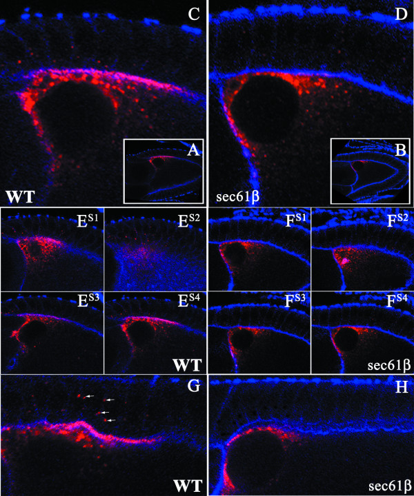
Localization of Gurken Protein in Stage 10 Egg Chambers of Wild type and sec61βP1 Germline Clones. Drosophila egg chambers of the indicated genotype, wild type, (WT) or egg chambers mutant for sec61β, (sec61β), stained for actin using labelled Phalloidin (blue) and Gurken (red). A-B) Low magnification images of wild type (A) and sec61β mutant egg chamber (B) at stage 10; Gurken appears at the anterior-dorsal position. C-D) Magnification of the anterior-dorsal region of wild type and sec61β mutant oocytes. Gurken co-localizes with actin in the wild type oocytes (C), where as the sec61β mutant oocytes do not show this co-localization (D). ES1-S4-FS1-S4) Optical sections of the egg chambers as shown in C and D, correspond to either wild type egg chambers (ES1-S4) or sec61β mutant egg chambers (FS1-S4). G-H) High magnification images of oocytes showing the region of the follicle cells from wild type (G) and sec61β mutant egg chambers (H). Gurken is seen in dot like structures within the follicle cells in the wild type egg chambers (indicated by arrows), these structures are absent in the mutant egg chambers.

