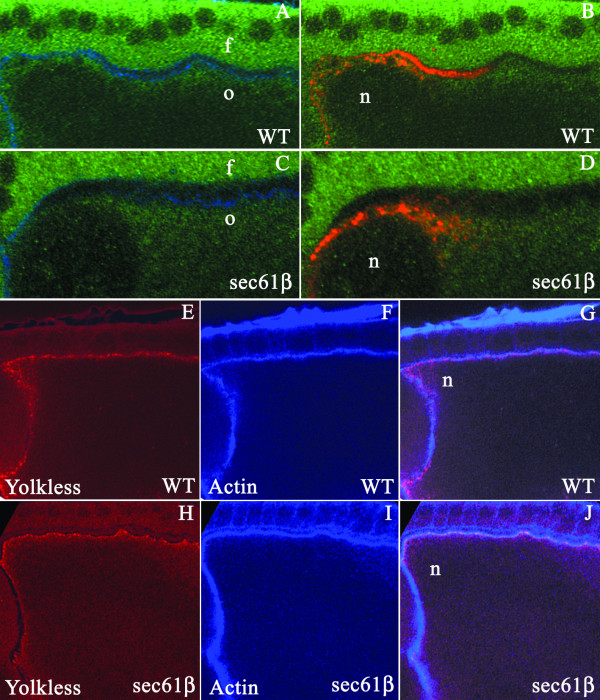
Figure 4.
Analysis of ER structure and function in sec61β mutant oocytes. A-D, Drosophila egg chambers of the indicated genotype, wild type, (WT) or egg chambers mutant for sec61β, (sec61β), stained for actin using labelled Phalloidin (blue), Gurken (red) and Boca (green). A and C) In oocytes from stage 10 egg chambers Boca stains a diffuse region below the plasma membrane that marks the ER in both the wild type (A) and the sec61βP1germline clones (C). B and D) Gurken staining is observed either along the plasma membrane or in distinct punctate structures (B) or Gurken is excluded from the plasma membrane and is present exclusively in the cytoplam (D). f, indicates the follicle cells, o, the oocyte and n, the oocyte nucleus. E-J, Drosophila egg chambers of the indicated genotype, wild type, (WT) or egg chambers mutant for sec61β, (sec61β), stained for actin using labelled Phalloidin (blue) and Yolkless (red). In wild type egg chambers (E) or egg chambers mutant for sec61β (H) Yl is at a peripheral location in the oocyte. Co-staining with phalloidin (F and I) shows that both in wild type oocytes and oocytes from the germline clones Yl co-localizes at the plasma membrane (G and J). n indicates the position of the nucleus.

