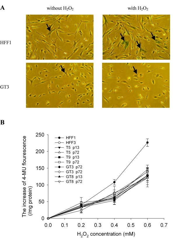
Figure 5.
H2O2-induced senescence in G6PD-deficient fibroblasts. A. Cells were treated with 350 μM H2O2 for 1.5 h and then cultured for another 72 h before staining for senescence-associated β-galactosidase (SA-β-Gal) activity. Representative results for HFF1 and GT3 cells with or without H2O2 treatment are shown in the right and left panels, respectively. Examples of cells that are positive for SA-β-Gal staining are indicated by arrow. In the quantification, any cell that has detectable green staining was scored as positive for SA-β-Gal. B. Cells were treated with different concentrations of H2O2 for 1.5 h and then cultured for another 72 h. Quantification of premature senescence was determined by the rate of conversion of 4-methylumbellliferyl-β-D-galactopyranoside (MUG) to fluorescent product 4-methylumbelliferone (4-MU) as described in M & M. Both the early passage (p13) and late passage (p72) cells of T5, T9, GT3 and GT8 were included in this study. Data are the means ± SD from 4 independent experiments.

