Fig. 3.
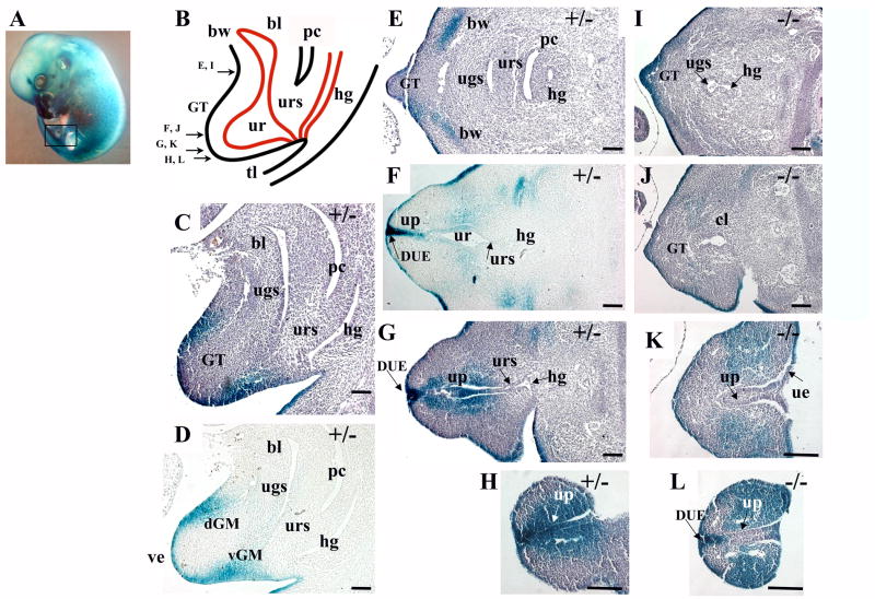
Bmp7 expression and null phenotype in the urogenital system at E13.5. (A) Bmp7lacZ/+ embryo at E13.5 stained with X-gal, left hindlimb is removed, the area of the sections is shown in a frame. (B) Schematic depiction of the urogenital area at E13.5. The genital tubercle (GT), cloaca (cl), urorectal septum (urs), bladder (bl), urethra (ur), hindgut (hg), peritoneal cavity (pc), body wall (bw) and tail (tl) are indicated. Arrows indicate planes of transverse sections in (E-L). (C, D) Sagittal sections of a Bmp7lacZ/+ embryo show Bmp7 expression in the ventral (vGM) and dorsal (dGM) genital mesenchyme. (E-H) Transverse sections of Bmp7lacZ/+ embryo show Bmp7 expression in the lateral mesenchyme of the body wall (E), in the epithelium of the urethra (ur) and the urethral plate (up) (F-H), and the adjacent genital mesenchyme (F-H). Strong LacZ activity was observed in the distal urethral epithelium (DUE) at the tip of the GT (F, G). (I-L) In the null, LacZ activity was lost in the medial (L) and proximal (K) parts of the urethral plate, but maintained in the DUE (L and G). In the null, LacZ-positive genital mesenchyme occupied a broad lateral domain (L). Scale bar: 100 μm.

