
Figure 2. Cardiac defect in SM22α-Cre;R26R;Bmpr1aflox/flox embryos.
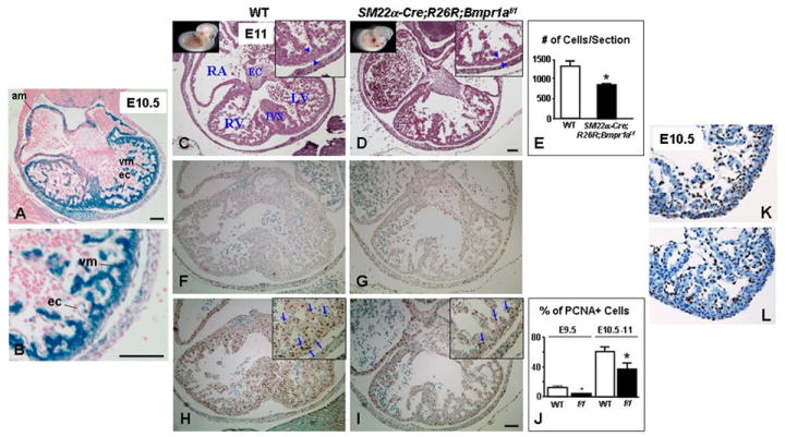
(A) shows blue staining (Cre activity) in the atrial (am) and ventricular (vm) myocytes but not in endocardial cells (ec) by whole-mount LacZ staining in E10.5 SM22α-Cre;R26R;Bmpr1aflox/flox embryo. B is an enlargement of A. Panels C, D, F, G, H and I are consecutive transverse sections of WT (C, F, H) and SM22α-Cre;R26R;Bmpr1aflox/flox (D, G, I) hearts taken at the same level from viable E11 embryos. H&E staining shows thinning of the ventricular wall (blue arrowheads) in the flox/flox mutant heart (D) vs. WT (C). E represents a numerical assessment of hematoxylin stained nuclei per ventricular section of E10.5-E11 WT (white bars) and SM22α-Cre;R26R;Bmpr1aflox/flox mutants (black bars). Bars represent mean±s.e.m. (n=3). *P<0.05. Apoptosis was infrequent in ventricular sections of E11 SM22α-Cre;R26R;Bmpr1aflox/flox mutant (G) and WT (F) by TUNEL immunostaining. However, fewer PCNA positive cells (brown) were observed in the mutant ventricles (I, blue arrows) compared to the WT (H, blue arrows). J represents a numerical assessment of percent of PCNA positive over total number of ventricular cells in heart sections of WT (white bars) and flox/flox mutant (black bars) embryos at E9.5 and E10.5-E11. Bars represent mean±s.e.m. (n=3-4). *P<0.05. Photomicrographs in K and L show a representative comparable p57kip2 immunostaining in E10.5 WT and flox/flox (f/f) mutant heart sections, respectively. Corner views in C, D, H, and I are higher magnifications. RA, RV, and LV denote right atrium and right and left ventricle, respectively. EC and IVS denote endocardial cushions and interventricular septum, respectively. Panels depicting WT and their corresponding mutants have the same magnification. Bars in A, B, D, and I=100 μm. F-I have the same magnification.