
Figure 3. Dilated large vessels with reduced VSMC in SM22α-Cre;R26R;Bmpr1aflox/flox embryos.
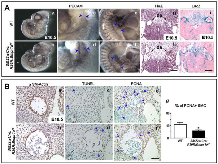
A. Whole-mount PECAM staining of E10.5 embryos (a,b) shows a dilated aorta in the SM22α-Cre;R26R;Bmpr1aflox/flox compared to WT embryo (d vs. c, arrowheads). Panels c and d are high magnifications of the framed areas in a and b, respectively. Dilatation of large abdominal vessels with frequent interconnections are seen in the flox/flox mutants vs. WT (f vs. e, arrowheads). H&E stained transverse sections show dilatation of the mutant aortae (h) compared to WT (g). Poor investment of blue SM22α expressing VSMC around the mutant aortae are noted in sections of whole-mount LacZ stained mutants (j) vs. WT (i) embryo. Sections h and j are at the same level as g and i, respectively. Panels depicting WT and mutants were acquired at the same magnification. Bars in h and j =100 μm. B. Alpha SM-actin staining (brown) shows poor investment of SMC in the dilated aortic wall of SM22α-Cre;R26R;Bmpr1aflox/flox (b) vs. WT (a). TUNEL staining (blue arrows) revealed similar occasional apoptotic cells surrounding the dilated aorta in the flox/flox mutant (d) and WT (c). However, reduced PCNA staining (blue arrows) was seen in VSMC of the dilated aorta and in the neighboring mesenchymal cells in the flox/flox (f) vs. WT (e). Panels a, c, and e are similar consecutive sections in the WT embryo as b, d, and f in the flox/flox mutant embryo. Panels a-f have the same magnification. Bar in f=50 μm. (g) Quantification of percent of PCNA positive over total number of SMC in the vessel walls in dorsal aortae of E10.5-11 WT (white bars) and SM22α-Cre;R26R;Bmpr1aflox/flox mutants (black bars). Bars represent mean±s.e.m. (n=4). *P<0.05.