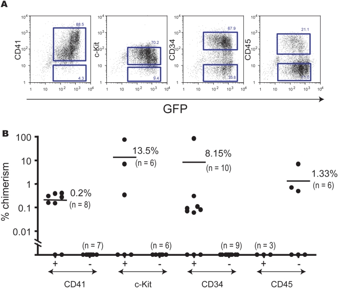
Figure 4. Surface markers of embryonic HSCs generated from EB6 cells in vitro.
(A) CD41+ EB6 cells were co-cultured with OP9 cells in the absence of Dox for 4 days. Cells collected from the co-cultures were analyzed by flow cytometry. HOXB4-expressing cells were detected by GFP expression. Data show the expression of CD41, c-Kit, CD34, and CD45 in GFP+ cells. The sorting gates for CD41− or CD41+ cells, c-Kit− or c-Kit+ cells, CD34− or CD34+ cells, and CD45− or CD45+ cells are shown as squares. (B) Co-cultured cells were fractionated based on expression of CD41, c-Kit, CD34, and CD45, and were transplanted into lethally irradiated mice. Recipient mice were analyzed 16 weeks after transplantation. Over 95% of reconstituted blood cells were of myeloid lineage in all cases (data not shown). Two independent experiments gave similar results. Data from one experiment are shown. See Table S2 for the number of transplanted cells for each subpopulation.

