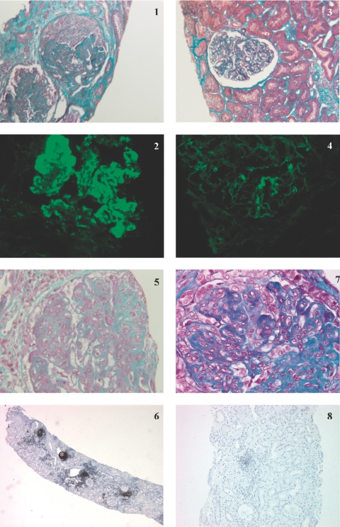
Figure 1.
Kidney biopsy specimens for patients 9 (panels 1 to 4) and 4 (panels 5 to 8) before and after rituximab (RTX). Patient 9: Kidney biopsy specimen obtained before the start of RTX showed (panel 1) global endocapillary and extracapillary proliferation on light microscopy study (Masson trichrome; original magnification: ×250), and (panel 2) granular Ig G deposits in the mesangium and along the basement membrane on immunofluorescence study (original magnification: ×400). A control kidney biopsy specimen obtained 5 mo after treatment with RTX showed (panel 3) very few active lesions on light microscopy study (Masson trichrome; original magnification: ×250) and (panel 4) a dramatic decrease in IgG glomerular immune deposits on immunofluorescence study (original magnification: ×400). Patient 4: Kidney biopsy specimen obtained before the start of RTX showed (panel 5) global endocapillary proliferation on light microscopy study (Masson trichrome; original magnification: ×400), and (panel 6) nodular CD20+ interstitial inflammatory infiltrate (*) on peroxydase immunohistochemical study (phenotyping of cells with anti-CD20 monoclonal antibodies; original magnification: ×20). A control kidney biopsy specimen obtained 2 mo after treatment with RTX showed (panel 7) global endocapillary proliferation and glomerular segmental fibrosis, (Masson trichrome; original magnification: ×400) contrasting with the disappearance of nodular CD20+ infiltrate on peroxydase immunohistochemical study (panel 8) (phenotyping of cells with anti-CD20 monoclonal antibodies; original magnification: ×40)

