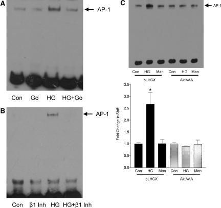
Figure 3.
PKC-β-Akt mediate activation of AP-1 by glucose. (A and B) MCs were treated with HG for 6 h in the presence or absence of the conventional PKC inhibitor Gö6976 (2 μM, 30 min; A) or specific PKC-β inhibitor (100 nM, 30 min; B). AP-1 activation, assessed by EMSA, was abrogated by both inhibitors. (C) AP-1 activation by HG was not observed in MCs overexpressing AktAAA as compared with empty vector pLHCX (n = 2; *P < 0.05 pLHCX HG versus others).

