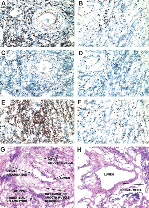
Figure 7.
Lack of dDC and worsening of chronic rejection. WT mice that received DT and MR1 were used as controls. Mice from both groups were sacrificed at day 100 after transplantation, and heart allografts were examined for cellular infiltrates; CD4+, CD8+, and Foxp3+ cells; and the severity of chronic rejection. Samples shown were recovered from DTR-GFP-DC donors treated with DT and MR1 (right) as well as from WT donors treated with DT and MR1 (left). (A, B, E, and F) The percentage of CD4+ and CD8+ infiltrating lymphocytes in the DTR-GFP-DC donors treated with DT (A and E, respectively) was significantly higher than those of WT donors (B and F). (A through D) Examination of the localization of Foxp3 expression revealed that whereas most infiltrating CD4+ cells overlap with Foxp3+ cells (B and D) in the WT donors, infiltrating CD4+ cells in DTR-GFP-DC donors treated with DT are predominantly non-Foxp3+ cells (A and C). (G and H) Interestingly, compared with WT donors (H), DTR-GFP-DC donors treated with DT showed substantially more severe chronic rejection, as is shown by increased cell numbers and a greater degree of smooth muscle necrosis/inflammation, and intimal thickening (G). Quantitative analysis of graft arteriolosclerosis demonstrated that luminal stenosis of coronary arteries in the DT-treated group (G) was significantly higher as compared with that of the WT group (H). The percentage of luminal stenosis for DT-treated and WT grafts was 85.0 ± 4.0 and 18.0 ± 3.5, respectively (n = 3 to 4 mice/group, P < 0.002).

