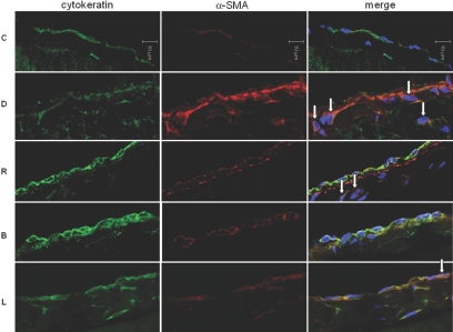
Figure 16.
Effect of BMP-7 on the number of submesothelial myofibroblast in an animal model of PD. Immunofluorescence microscopy stained for cytokeratin (green) and α-SMA (red) with nuclear counterstain (blue, DAPI) demonstrates an increase in submesothelial dual-stained cytokeratin- and α-SMA–positive myofibroblasts (arrow) in group D associated with a decrease in cytokeratin and an increase in α-SMA expression. Peritoneal rest decreases the number of these cells in group R, which is further decreased by Adv-BMP-7 transfection (group B). Magnification, ×640.

