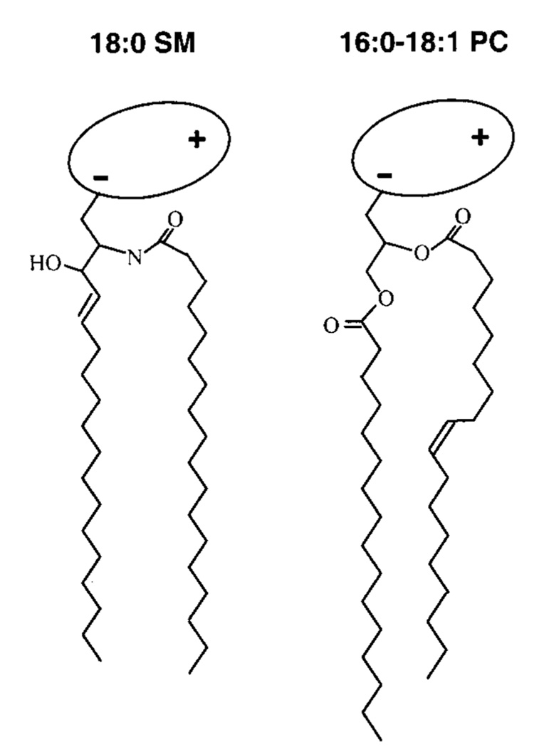
FIGURE 1.
Structural features of naturally predominant sphingomyelin (SM) and phosphatidylcholine (PC). For simplicity, the lipid hydrocarbon chains are depicted as fully extended trans rotamers. In reality, free rotation about the carbon–carbon single bonds provides numerous trans–gauche isomers in the fluid but not the gel state. The cis double bond depicted in the sn-2 chain of PC leads to a t+gt− kink. Rotation about nearby carbon-carbon bonds increases the average molecular cross-sectional area. In PC, the glycerol base is depicted in gray and the highly hydratable, zwitterionic phosphocholine headgroup is depicted as a shaded ellipse oriented roughly parallel to the bilayer surface. In SM, the phosphocholine headgroup is depicted similarly, and a stearoyl (18: 0) acyl chain, typical of bovine brain SM, is shown amide-linked to the 18-carbon sphingosine base, which contains a 4,5-trans double bond.

