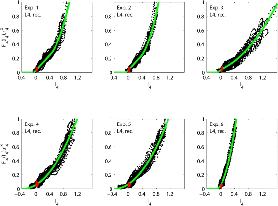
Figure 4. Fits of recurrent thalamocortical model.
Illustration of fits of recurrent thalamocortical model in Eq. (2) to
data from experiments 1–6. Each black dot corresponds to
the experimentally measured layer-4 firing rate  at a specific time point
  plotted against the model value of
  . The red dots are corresponding experimental data
points taken from the first 5 ms after stimulus onset (for all 27
stimuli). These data points show the activity prior to any
stimulus-evoked thalamic or cortical firing and correspond to
background activity. The solid green curve corresponds to the fitted
model activation function
  .
.

