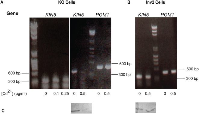
Figure 4. Optimization of KIN5 shRNA.
A. Degradation of KIN5 message after shRNA induction in KO cells using 0–0.5 µg/ml Cd2+. Above: RT-PCR products resolved on a 1% agarose gel. At Cd2+ concentrations lower than 0.5 µg/ml, KIN5 mRNA is stable for 24 h. After 24 h in 0.5 µg/ml Cd2+, KIN5 mRNA is dramatically decreased, while PGM1 is unaffected. B. Effect of 0.5 µg/ml Cd2+ on KIN5 and PGM1 messages in Inv2 cells. KIN5 and PGM1 mRNA levels remain unaffected after 24 h. DNA markers shown: lines indicate 600 and 300 bp. C. Effect of 0.5 µg/ml Cd2+ on Kin5 protein levels in KO and Inv2 cells. Corresponding KO (left) and Inv2 (right) cell homogenates 12 h post-induction at either 0 or 0.5 µg/ml Cd2+ and blotted with K5T1 Ab to Kin5. While the Kin5 protein is severely knocked down in the KO cells upon shRNA induction, Kin5 levels remain unaffected in Inv2 cells under similar conditions.

