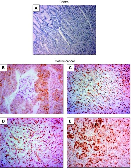
Figure 1.
Expression pattern of HIF-1α in human gastric cancer tissues. Paraffin sections were pretreated as described in Materials and Methods, and HIF-1α was visualised by means of immunohistochemistry. (A) Negative control staining. (B–E) Expression of HIF-1α in established human gastric cancers, showing that the vast majority of tumour cells were positively stained for HIF-1α over nuclei. Magnification × 100 (A and B) and × 200 (C–E).

