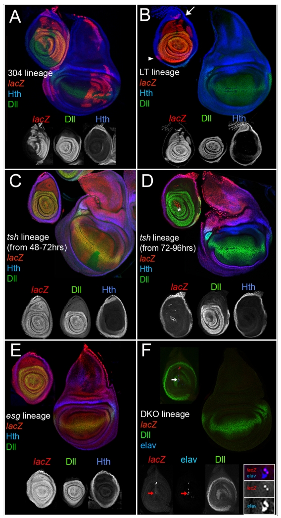
Fig. 2.
Lineage analyses of genes active in the ventral limb primordia. All discs except for those in F were stained for the lineage marker (red), Dll (green, subset of telopodite), and Hth (blue, coxopodite); see Materials and methods for details. (A) The progeny of cells in which Dll304 was active contribute to both dorsal (wings and halteres) and ventral (legs, both coxopodite and telopodite) thoracic limbs. Although individual wing discs show labeling in only a subset of the disc, labeled cells can contribute to any part of the disc. (B) The progeny of cells in which LT was active generate the telopodite of the leg. Expression in the dorsal coxopodite (arrow) may be due to the imperfection of the LT-Gal4 driver. The arrowhead marks a clone in the trochanter region. (C,D) The progeny of cells that expressed tsh become more restricted over time. Restricting tsh-Gal4 activity to the beginning of second instar (48-72 hours AEL) results in the labeling of both the coxopodite and telopodite (C). Allowing tsh-Gal4 to be active beginning at third instar (72-96 hours AEL) results in the labeling of only the coxopodite (D). The asterisk in D indicates lacZ-positive adepithelial cells that are not part of the disc epithelium. (E) The progeny of esg-expressing cells adopt both wing and leg (coxopodite and telopodite) fates. (F) The progeny of the cells in which DKO was active (red) occasionally contribute to larval neurons that co-express Elav (blue, arrow). All lacZ-positive cells express elav but not all elav cells are lacZ positive (see inset).

