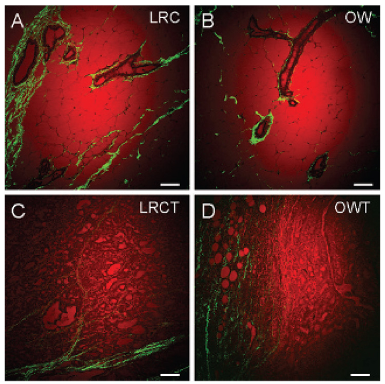
Figure 4.
Coherent anti-Stokes Raman scattering imaging of lipid (red) and second harmonic generation imaging of collagen fibrils (green) of standard histologic tissue sections. Histology of the mammary gland of (A) a lean rat chow rat (LRC), (B) an obese Western rat (OW), and the mammary tumors of (C) a lean chow tumor rat (LRCT) and (D) an obese Western tumor rat (OWT). Images taken with a 20× air objective. Scale bars = 75 µm.

