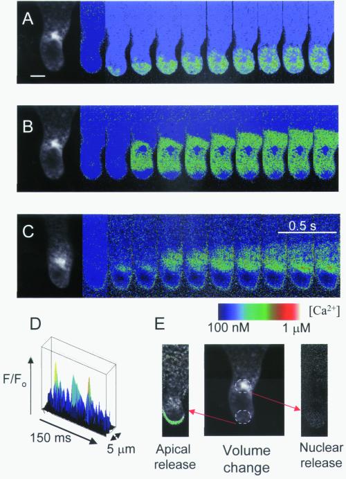
Figure 4.
Patterns of Ca2+ elevation in response to localized photorelease of caged Ins(1,4,5)P3. (A) Photorelease of Ins(1,4,5)P3 from a 10-μm diameter region at the rhizoid apex (E) caused Ca2+ elevation that propagated in a subapical direction but was generally (60% of cells, n = 23) restricted to the apical 40 μm. (B) Ins(1,4,5)P3 release in the nuclear region induced a Ca2+ wave that propagated bidirectionally and did not permeate the nucleoplasm (8 out of 14 cells). (C) In a further six cells, Ca2+ release in the nuclear region gave an initial Ca2+ increase in the nucleoplasm followed by perinuclear Ca2+ waves. (D) Elemental Ca2+ elevations (>2 SD above the mean background) could be observed during propagation of Ins(1,4,5)P3-induced Ca2+ waves in apical and subapical (not shown) regions. (E) Representative volume decrease in response to nuclear or apical Ins(1,4,5)P3-induced Ca2+ elevations. The green area indicates volume decrease in a representative cell after release of caged Ins (1,4,5)P3 in the apical rhizoid regions (n = 5). No volume decrease could be observed following Ins(1,4,5)P3 release in nuclear regions. (Bar = 15 μm.)

