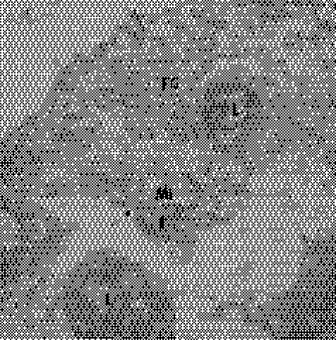
Figure 8 Electron microscopy of the heart of a patient who died of acute Chagas disease. Notice lymphocytes (L) invading the cytoplasm of a cardiac fibre (FC), adhering to the microfilaments (Mi) and concomitant lyses (courtesy of Dr Washington Luiz Tafuri, Faculdade de Medicina da Universidade Federal de Minas Gerais). Magnification ×7000.

An official website of the United States government
Here's how you know
Official websites use .gov
A
.gov website belongs to an official
government organization in the United States.
Secure .gov websites use HTTPS
A lock (
) or https:// means you've safely
connected to the .gov website. Share sensitive
information only on official, secure websites.
