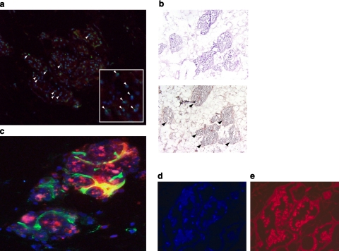
Figure 6.
Engraftment and proliferation of MAPCs and MSCs onto EMBs. a) Following infusion, gender-mismatched DiI-labeled MAPCs (red) can be readily identified by FISH (green, Y chromosome, white arrowheads; inset ×400). b) BrdU staining of MAPC clusters demonstrates proliferation of stem cells following engraftment (black arrowheads). c) MAPC clusters marked with DiI (red) and dapI (blue) staining stimulate angiogenesis or differentiate into functional neovessels (vasculogenesis) seen with lectin (green) perfusion within the EMB. d) Human MSCs engrafted onto EMBs and formed viable clusters, as seen on dapI staining. e) PKH26-fluorescent labeling.

