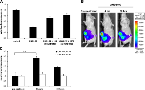
Figure 9.
In vivo imaging of CXCR4 receptor dimers. A) Type 231 cells stably transduced with CXCR4 dimerization reporters were incubated in cell culture with 1 μg/ml CXCL12 for 15 min. AMD3100 or vehicle control then was added to CXCL12-containing medium for 4 h before quantifying bioluminescence in living cells (n=4/condition). Data are presented as mean ± se bioluminescence relative to values measured in cells not treated with CXCL12 or AMD3100 (representative of 2 independent experiments). B, C) Type 231 CXCR4 homodimer or CXCR4-CXCR7 heterodimer cells were coimplanted with CXCL12-producing fibroblasts into mammary fat pads of nude mice. B) Representative bioluminescence images of the same mouse prior to treatment with 7.5 mg/kg AMD3100 i.p. and then 4 and 30 h later. CXCR4 homodimer tumors are on the right side of the mouse; CXCR4-CXCR7 heterodimers are on the left. Bioluminescence is shown as a pseudocolor scale, with red the highest and blue the lowest levels of photons. C) Quantified data from in vivo imaging experiments for CXCR4 and CXCR4-CXCR7 tumors (n=4/tumor type). **P < 0.01.

