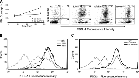
Figure 4.
PBL counts and PSGL-1 expression in EIU animals with anti-PSGL-1-neutralizing mAb. A) At 6 h after LPS injection, mice were treated with PSGL-1-neutralizing mAb (4RA10) or isotype control. The animal blood was harvested at 30, 120, and 180 min after antibody treatment, and the PBL counts were obtained. In parallel, leukocytes were evaluated for their PSGL-1 surface expression through indirect immunofluorescence. B) PSGL-1 surface expression on PBLs from EIU mice 6 h after LPS injection at 30, 120, and 180 min after antibody treatment. PBLs from animals 120 and 180 min after PSGL-1 blockade showed higher expression of PSGL-1 compared with the cells obtained 30 min after mAb treatment. C) Increased PSGL-1 surface expression on PBLs from EIU mice 6 h after simultaneous administration of PSGL-1-neutralizing mAb (4RA10) and LPS, compared with 4RA10-treated controls. Each curve is representative of 3 independent experiments.

