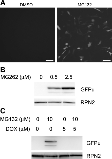
Fig. 1.
Doxorubicin (Dox) enhances GFPu degradation in 3T3 cells. A: epifluorescence micrographs of GFPu stably transfected 3T3 cells 6 h after treatment with a proteasome inhibitor (MG132; 10 μM) or vehicle control (DMSO). GFPu is a green fluorescent protein modified with carboxyl fusion of degron CL1. Scale bar = 10 μm. B: representative Western blot images showing dose-dependent increases in GFPu by MG262. C: Western blot images showing that MG132-induced GFPu accumulation was abolished by cotreatment with Dox. In both A and B, the same membranes were reprobed for RPN2 (a subunit of the 19S proteasome) to illustrate that the amount of total proteins loaded to the lanes absent of GFPu signals was not less than the lanes where GFPu was detected.

