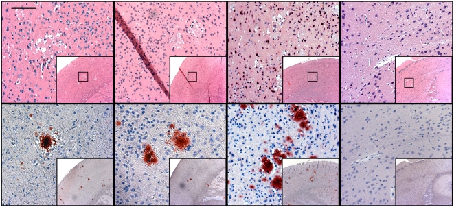
Figure 1. Spongiform degeneration and PrPCWD identified by histopathology and immunohistochemistry.
Vacuolated neurons and spongiform degeneration of the neuropil characteristic of a TSE is evident on H&E staining, with the colocalization of PrPCWD specific immunostaining of florid plaques in the cortices of mice inoculated with positive control inoculum and concentrated urine and saliva from CWD-infected cervids. Negative control mice showed no evidence of spongiform degeneration or PrPCWD immunostaining. HRP-conjugated BAR-224 was used as a primary antibody. (Measure bar, 50 µm).

