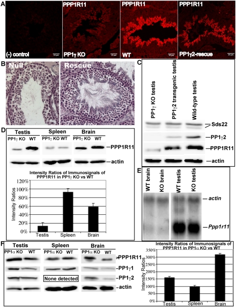
Figure 9. Testicular phenotypes related to the expression of PP1γ2.
A. Immunofluorescence of PP1γ-null (KO), wild-type (WT), and PP1γ2-rescue testis sections probed with anti-PPP1R11 antibody. Negative (−) control = no primary antibody. B. Histological analysis of hematoxalin-stained testis sections from PP1γ-null (Null) and PP1γ2-rescue (Rescue) mice demonstrating an antiapoptotic effect of PP1γ2. C. Western blot of testis protein extracts from PP1γ-null (left lane), PP1γ2-rescue (center lane), and wild-type (right lane) animals probed with anti-Sds22, anti-PP1γ2, anti-PPP1R11, and anti-actin antibodies. D. Upper panel: PPP1R11 level in PP1γ-null spleen is not significantly diminished compared to its level in wild-type spleen, but is significantly diminished in tissues normally expressing PP1γ2 (testis and brain); Lower panel: histogram of immunosignal ratios of PPP1R11 in PP1γ-null vs wild-type tissues. E. Northern blot analysis showing that Ppp1r11 mRNA levels are comparable in PP1γ-null and wild-type brain and testis, respectively. Actin mRNA levels are used as gel loading control. F. Left panel: PP1γ isoform levels are increased and PPP1R11 levels rise dramatically in PP1α-null vs wild-type testis and brain, but PP1γ1 and PPP1R11 levels in PP1α-null spleen are unchanged; Right panel: histogram of immunosignal ratios of PPP1R11 in PP1α-null vs wild-type tissues.

