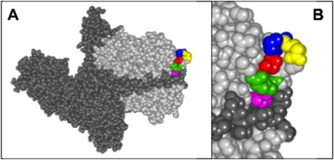
Figure 3. Location of the QPDRS motif within the BoNT/A holotoxin.
A. Space-filled diagram of the three-dimensional model of the complete BoNT/A holotoxin showing the heavy (dark gray) and light (light gray) chains. QPDRS motif is the colored region within the light chain. B. Enlarged view of Q139 (red), P140 (blue), D141 (yellow), R145 (green) and S146 (pink) [7].

