Figure 7.
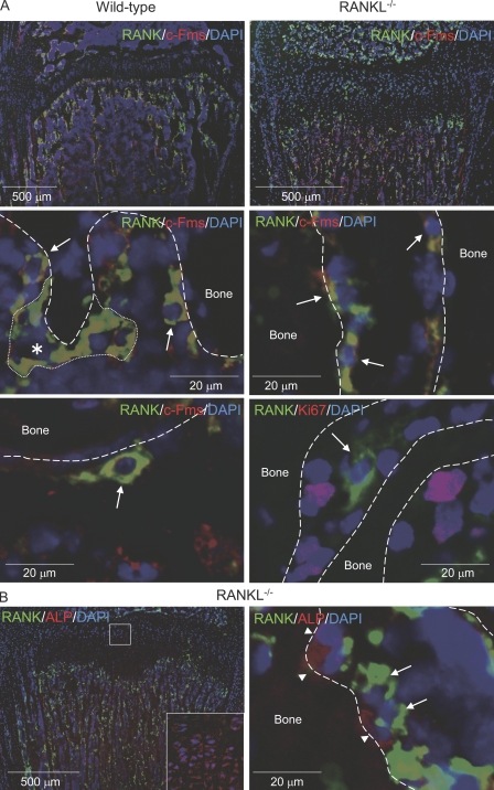
Localization of QuOPs in bone. (A) Localization of c-Fms+/RANK+ and Ki67+ cells. Tibiae were recovered from 7-wk-old wild-type and 3-wk-old RANKL−/− mice. Sections of tibiae were prepared and subjected to double staining of RANK (green) and c-Fms (red; top and middle). Nuclei were labeled with DAPI (blue). Top panels show low power views of the specimens, and middle and bottom panels show high power views. (middle) The asterisk indicates a multinucleated osteoclast, which is surrounded by a small dotted line, and arrows indicate mononuclear cells double positive for RANK and c-Fms (yellow). Tibiae were also recovered from 7-wk-old wild-type mice pretreated with 5-FU for 6 d. (bottom left) Sections of tibiae from 5-FU–treated mice were stained for RANK (green), c-Fms (red), and DAPI (blue). The arrow indicates cells double positive for c-Fms and RANK in 5-FU–treated mice. (bottom right) Sections of tibiae from RANKL−/− mice were also stained for RANK (green), Ki67 (red), and DAPI (blue). The arrow indicates a RANK+ and Ki67− cell. Bones are indicated by dashed lines. (B) Localization of RANK+ cells and ALP+ cells. Sections of tibiae from RANKL−/− mice were subjected to staining of RANK (green), ALP (red), and DAPI (blue). Arrows and arrowheads indicate RANK+ and ALP+ cells, respectively. The inset shows a high power view of the boxed region.

