Figure 6.
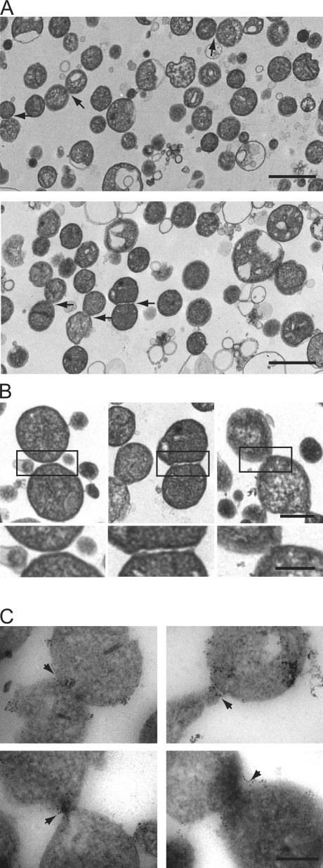
Identification of mitochondrial outer membrane fusion intermediates. (A) Analysis of outer membrane tethering. Representative electron micrograph fields of wild-type (top) and ugo1-2 (bottom) mitochondria subjected to in vitro outer membrane fusion at the nonpermissive temperature are shown. Tethered mitochondria are indicated by arrows. (B) Representative images of outer membrane–tethered mitochondria from analysis described in A. The boxed regions are enlarged below. Bars: (top) 0.5 µm; and (bottom) 0.25 µm. (C) Immuno-EM analysis of Fzo1 localization in wild-type mitochondria subjected to S1 in vitro fusion conditions in the presence of the nonhydrolysable GTP analogue GMP-PCP. Arrows indicate the interface at the outer membrane formed by two mitochondria, decorated Fzo1-specific immunogold particles. (A and C) Bars, 0.5 µm.

