Figure 2.
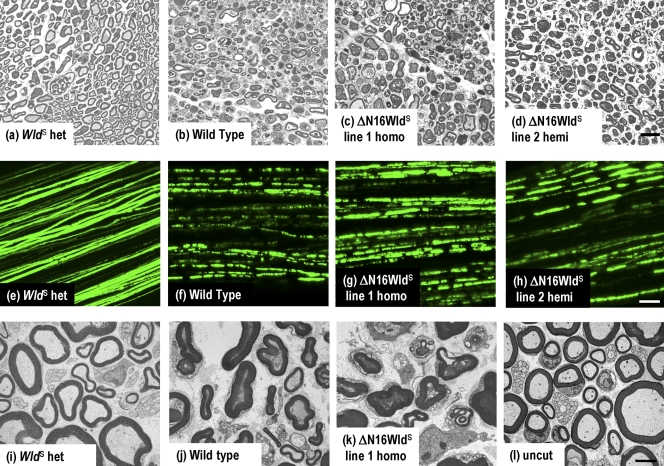
Rapid Wallerian degeneration in ΔN16WldS Tg mice. (a–d) Semithin sections of distal sciatic nerve 5 d after lesion. (a) Axons are well preserved in WldS heterozygotes, with intact myelin sheaths, uniform and regularly spaced cytoskeleton, and normal-shaped mitochondria. (b) Wild-type axons are degenerated, with collapsed myelin and disorganized or vacuolized cytoskeleton. (c and d) ΔN16WldS line 1 homozygous and line 2 hemizygous nerves are indistinguishable from wild type. (e–h) Tibial nerves from mice crossed to YFP-H show rapid loss of axon continuity in ΔN16WldS. (e) WldS heterozygotes 3 d after lesion show axon continuity. (f–h) In contrast, all lesioned wild-type and ΔN16WldS line 1 and line 2 axons lose continuity within 72 h. (i–l) Transmission electron microscopy of distal sciatic nerve 3 d after lesion. (i) Myelinated and unmyelinated axons are well preserved in WldS heterozygotes, which are indistinguishable from unlesioned nerves (l). (j) In wild type, myelin collapsed to form ovoids, the cytoskeleton is floccular or absent, and mitochondria are swollen or absent. (k) Nerves from ΔN16WldS line 1 homozygotes are indistinguishable from wild types. Bars: (a–d) 20 µm; (e–h) 50 µm; (i–l) 2 µm.

