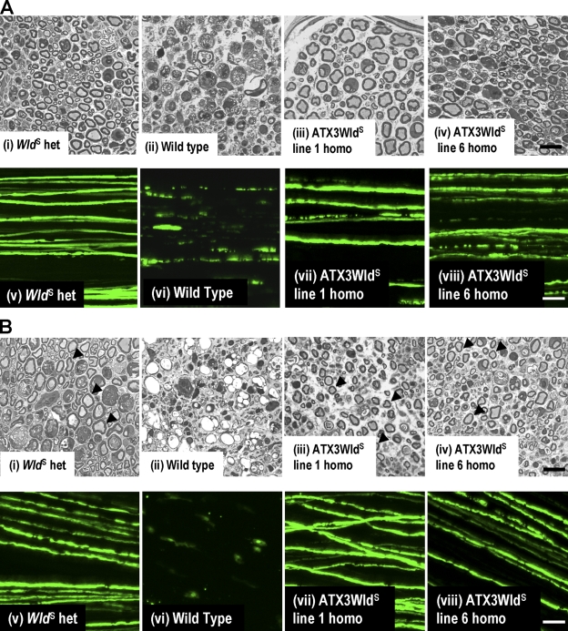
Figure 3.
ATX3WldS Tg mice show a WldS phenotype. (A, i–iv) Semithin sections of distal sciatic nerve 5 d after lesion. (i and ii) Most axons are well preserved in WldS heterozygotes (i), but wild-type nerves (ii) are completely degenerated. (iii and iv) Nerves from ATX3WldS line 1 and line 6 show preservation similar to WldS heterozygotes. (v–viii) Confocal images of tibial nerve 5 d after sciatic lesion from mice crossed to YFP-H. (v) WldS heterozygotes maintain axon continuity. (vi) All wild-type axons are highly fragmented. (vii and viii) Many lesioned ATX3WldS axons maintain continuity. (B, i, iii, and iv) Semithin sections of distal sciatic nerve show preserved axons (arrowheads) 14 d after lesion in WldS heterozygotes (i) and ATX3WldS (iii and iv). (ii) Wild-type axons are completely degenerated and highly vacuolized. (v–viii) Confocal images of tibial nerves 14 d after sciatic nerve lesion in mice crossed to YFP-H. (v) WldS axons maintain continuity in contrast to the few remnants of the fragmented wild-type axons (vi). (vii and viii) Many axons in ATX3WldS were also continuous at this stringent time point. Bars: (i–iv) 20 µm; (v–viii) 50 µm.

