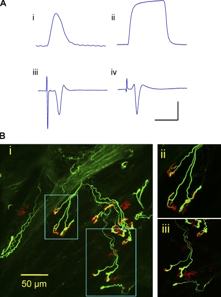
Figure 4.
Functional motor innervation. (A) ATX3WldS mice show robust preservation of neuromuscular function 3 d after nerve lesion. (i and ii) Isometric single twitch (i) and 20-Hz tetanic tension (ii) responses of isolated FDB to tibial nerve stimulation. (iii and iv) Averaged (16 sweeps) extracellular evoked EMG responses to stimulation of 3-d axotomized FDB from ATX3WldS (iii) and WldS (iv). The x axis represents milliseconds, and the y axis represents millinewtons or microvolts. Bars: (i) 200 ms and 2 mN; (ii) 700 ms and 5 mN; (iii) 6 ms and 200 µV; (iv) 12 ms and 200 µV. (B) Confocal microscopy of axotomized FDB from an ATX3WldS line 5 mouse in which physiological recordings indicated robust preservation of neuromuscular function. Most motor endplates (TRITC–α-bungarotoxin staining; red) were innervated by neurofilament-positive axon collaterals and motor nerve terminals (green), but ∼20% of endplates were unoccupied. (i) Low power z-series projection. (ii and iii) Higher magnification images correspond to the boxed areas in panel i.

