Figure 5.
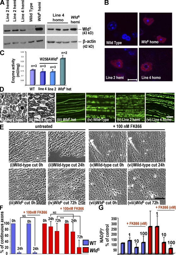
Rapid Wallerian degeneration in W258AWldS Tg mice. (A) Brain Western blots from W258AWldS Tg, WldS, and wild-type mice probed with Wld18. W258AWldS lines 2 and 4 express a 43-kD band, which is absent in wild type. (B) Wld18 immunofluorescence (red) of lumbar spinal cord. Motor neuron nuclear signal strength and distribution in W258AWldS lines match WldS heterozygotes. Identical laser intensities and camera settings were used for each image. (C) Nmnat1 activity is unaltered in the W258AWldS brain. (D, i and ii) Semithin sections of W258AWldS distal sciatic nerve 72 h after lesion. Axons are degenerated, similar to wild-type or ΔN16WldS axons (Fig 2). (iii–vi) In mice crossed to YFP-H, tibial nerve axons lose continuity within 72 h of sciatic lesion, except in WldS (iii). (E) SCG explants untreated (i–iv) or treated (v–viii) with 100 nM FK866 for 72 h and then cut. Unlike wild-type axons (i, ii, v, and vi), untreated WldS axons are intact 72 h after being cut (iv) but when treated with FK866 are more degenerated (viii). (F) Quantification of continuous neurites (or neurite bundles). ***, P < 0.0001 (one-way analysis of variance followed by Bonferroni post-hoc test); n = 8 (wild type [WT]) and 9 (WldS). (G) NAD+ or NADP+ levels in wild-type and WldS explants ± FK866 at 1–100 nM. Mean of three different experiments. (C, F, and G) Mean ± SD. Bars: (B, D, i and ii, and E) 10 µm; (D, iii–vi) 100 µm.

