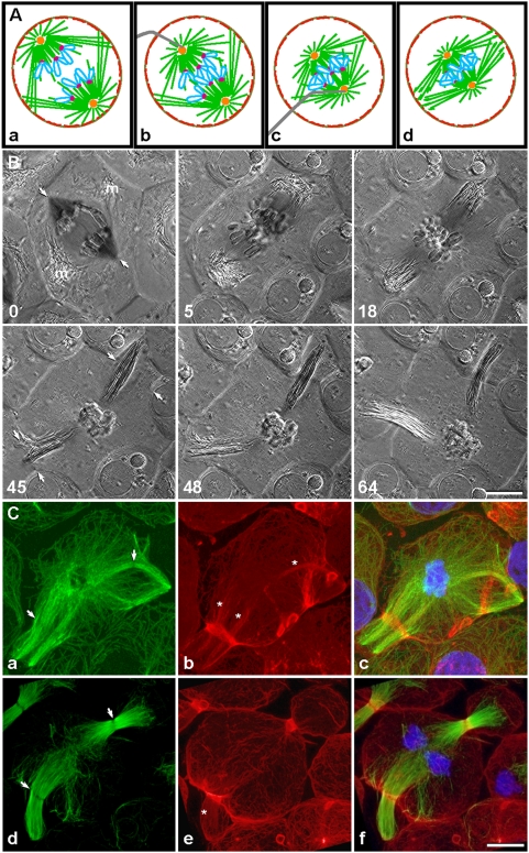
Figure 1. Mechanically induced reorganization of microtubules mediated actin redistribution into ectopic contractile rings.
(A, a–d) The diagram shows how an anaphase spindle can be mechanically collapsed by pushing the spindle poles together, using a micromanipulation needle. The procedure results in ectopic cleavage in two locations, as shown in (B). Microtubules, green; chromosomes, blue; kinetochores, magenta; centrosomes, orange; cortical actin, red; needle, gray. (B) Polarization microscopy sequence of ectopic cleavage, with time shown in minutes. Following anaphase onset (0 min, arrows show direction of impending collapse; m, spindle-associated mitochondria; also see Video S1), the spindle was collapsed with a microneedle, resulting in lateral growth of microtubules that dislodged the mitochondria (5 min). On opposite sides of the cell, these microtubules reorganized into two lateral spindles, while the displaced mitochondria appeared to rebundle with the microtubules (18 min). Cleavage furrows initiated simultaneously around both of the continuously reorganizing lateral spindles (45 min, arrows). Ultimately, each furrow ingressed around the approximate midpoint of its spindle (48 min), producing two anucleate membrane pockets (64 min). (C) Distribution of microtubules (green), actin filaments (red), and chromosomes (blue) in cells fixed at furrow initiation (a–c) and ingression (d–f) showed that each lateral spindle was in fact an independent bipolar spindle complete with a midzone (a and d, arrows). Bands of actin filaments (b and e), presumably contractile rings, encircled the midzone regions, while another subset of actin filaments (marked by asterisks) appeared to colocalize with spindle microtubules. Remnants of contractile rings from previous cell cleavages, i.e., “cell division scars”, were also present (b and e; small circles or ovals at cortex). Bars, 10 µm.

