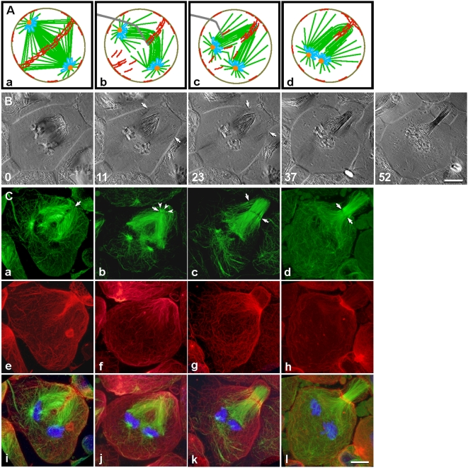
Figure 2. Spindle folding generated a concentrated source of actin for building a new contractile ring.
(A) The central spindle can be folded using a microneedle, before furrow formation. Color scheme, as in Fig. 1A. (B) Polarization microscopy sequence (Video S2). After folding (0 min), the mechanically generated monopolar spindle reorganized into a bipolar spindle. Microtubules from the pair of spindle poles radiated toward the cell cortex (11 min, arrows) where a furrow initiated (23 min, arrows). The furrow ultimately ingressed at the equator of the spindle (37–52 min), after realignment of the spindle midzone. (C) Distribution of microtubules (green), actin filaments (red), and chromosomes (blue) in cells fixed at stages similar to those in (B). Upon folding of the spindle by micromanipulation, the deformed midzone (a, arrow) and remnants of the contractile ring (e and i) remained relatively organized. As central spindle microtubules reorganized, the original midzone disappeared (b). Concurrently, actin filaments dispersed from the disintegrating ring, oriented in apparent alignment with central spindle microtubules (f and j). Meanwhile, a few very short microtubule bundles (b, arrowheads) emerged at the spindle tip and initiated formation of a new midzone, transverse to the new spindle axis (b, arrow). Around the time of furrow initiation, reorganization of the elongated bundles created a new “pole” (c), thereby establishing a bipolar spindle with a broad midzone (c, arrows). Actin filaments appeared to disassociate from spindle microtubules as they entered the new contractile ring (g and k). The furrow ingressed as microtubules at the distal pole elongated (c–d, green), shifting the midzone (c–d, arrows) and the contractile ring (g–h and k–l) toward the spindle equator. Bars, 10 µm.

