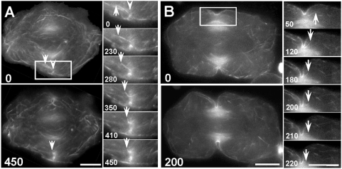
Figure 3. Incorporation of actin filaments into the contractile ring during furrow induction and ingression.
(A) Surface view of induction. Actin filaments were labeled by microinjection with Alexa 488-phalloidin and imaged every 10 seconds. The large panels show overall actin redistribution over time. The small panels show the dynamics of a single actin filament (arrows), as seen in selected time-lapse images within the boxed region of interest (Video S3). Immediately prior to furrow initiation (0 sec image), some cortical actin filaments began to move toward the spindle equator where they coalesced into nascent bundles of the emerging contractile ring (arrowhead). The arrows mark the end of a filament of interest, discernible at this focal plane. The filament moved toward the equator, underwent a sharp change in trajectory, and disappeared into the emerging contractile ring. The unlabeled spindle was flanked by microtubule-associated, autofluorescent mitochondria (also present in (B)) that hampered visualization of actin filaments in the vicinity. Time in seconds. Bars, 10 µm. (B) Midplane view of ingression. Actin filaments were labeled and observed as described in (A) except imaged at the midplane of the cell to follow furrow ingression. In the large panels, both labeled actin filaments and autofluorescent mitochondria could be seen. During furrow ingression (arrowheads), actin filaments continued to move toward and incorporate into the contractile ring, presumably along spindle microtubules. The small panels (selected from Video S4; within the boxed region) show the dynamics of a single actin filament (arrows) moving into the ingressing contractile ring. Time in seconds. Bars, 10 µm.

