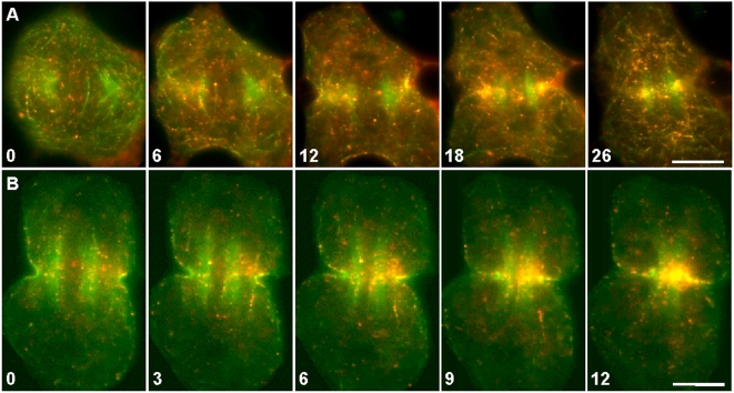
Figure 5. Redistribution of actin filaments labeled by Alexa 488-Phalloidin and Qdot 655-Phalloidin.
(A–B) Actin filaments (green) speckled with Qdots (red) continuously merged into the contractile ring during furrow induction (A; Video S6) and furrow ingression (B; Video S7). Occasionally, Qdot-decorated actin filaments could be seen moving away from the contractile ring (B, 3–9). The unlabeled spindles were flanked by a pair of green bundles that contained microtubule-associated, autofluorescent mitochondria. Time in minutes. Bar, 10 µm.

