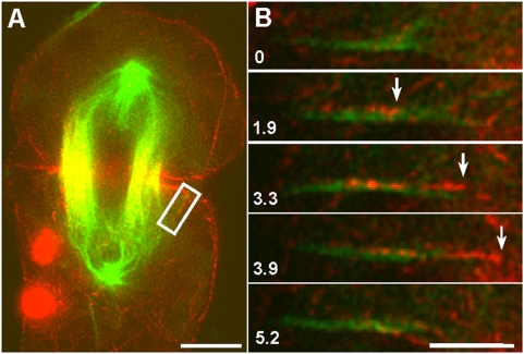
Figure 7. The movement of an actin filament along sparsely distributed spindle microtubules during furrow ingression.
For fluorescent speckle microscopy, microtubules (false-colored green) and actin filaments (false-colored red) were labeled by microinjection with Rhodamine-tubulin and a minute amount (∼0.2 µM) of Alexa 488-phalloidin, respectively. (A) A selected image from a time lapse series showing a dividing cell in which a single, clearly visible microtubule (boxed) happened to be isolated from the rest of the central spindle. Bar, 10 µm. (B) Sequential images of the boxed region, containing the cell cortex and the microtubule of interest, were enlarged and reoriented, with the microtubule horizontal and the cell cortex toward the right. A speckled actin filament (or bundle) appeared to move along a single microtubule toward the cortex. Bar, 5 µm.

