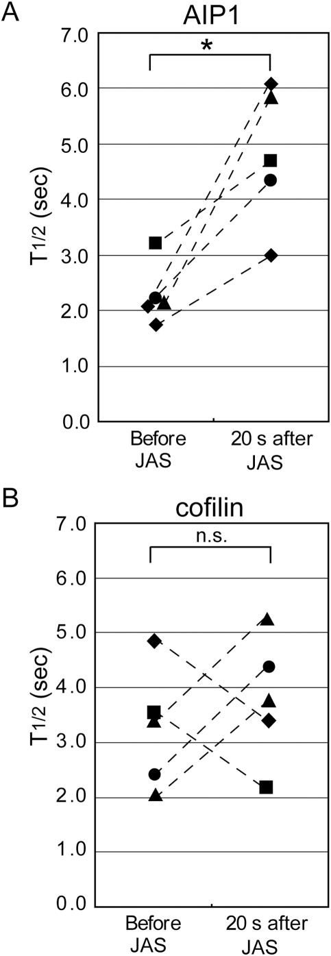
Figure 4. Dissociation of AIP1, but not of cofilin, slows by jaskplakinolide treatment; analogous to Jas-induced stabilization of capping protein.
Images of EGFF-AIP1 (A) or XAC2-EGFP (B) speckles in live XTC cells were acquired before and 20–40 s after treatment with 2 µM jasplakinolide. The decay rate of persistent single-molecule AIP1 or cofilin speckles before and 21–34 s after drug perfusion is compared in the same cells (connected by dashed lines) and expressed as half-life (n = 5 cells for each construct). *p = 0.012 (A), n.s.: not significant (B), paired t-test, two-tailed.

