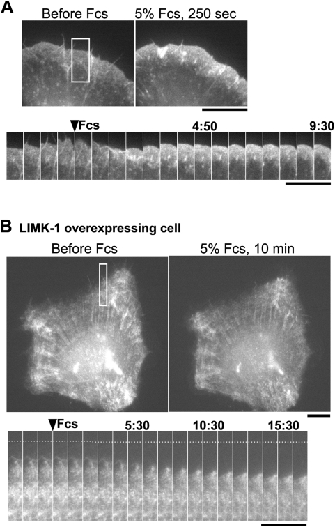
Figure 7. Inhibition of serum-induced lamellipod actin assembly by overexpression of hLIMK-1.
(A) XTC fibroblasts were serum-starved for ∼4 h after spreading on poly-L-lysine coated coverslips and then stimulated by 5% fetal calf serum (FCS). The upper panels show EGFP-actin before and 250 s after perfusion of L-15 medium containing 5% FCS. The lower panels show time-lapse images at 40 s intervals (square). The cell edge gradually extended after serum stimulation accompanied by a marked increase in EGFP-actin fluorescence in lamellipodia. (B) The upper panels show EGFP-actin images in an XTC cell overexpressing mRFP-hLIMK-1. The lower panels are time-lapse images of EGFP-actin at the intervals of 60 s (square). Upon 5% FCS stimulation, lamellipodial F-actin structure contracts toward the cell center accompanied by a slight increase in the density of EGFP-actin. However, the total amount of EGFP-actin fluorescence associated with the peripheral actin structures was not increased. Time is in minute:second. Bars, 10 µm.

