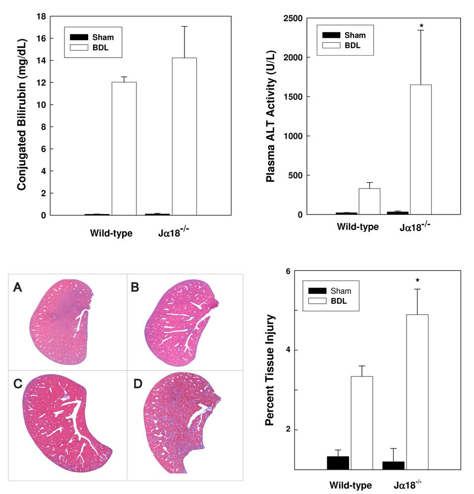
Figure 3. iNKT cells suppress cholestatic liver injury.
Sham operations or BDL were performed on groups of 6 wild-type and J α 18−/− mice. Plasma was collected on day 3 post-surgery; conjugated bilirubin (top panel, left) and ALT levels (top panel, right) were quantified [*significantly greater than other groups; P <0.05]. Livers dissected from wild-type (A/C) and Jα18−/− (B/D) mice on day 3 following sham operations (A/B) or BDL (C/D) were sectioned, stained with trichrome stain (bottom panel, left; 100-fold magnification) and subjected to photoimage analysis (bottom panel, right). Percent damaged area stained blue/total area ± SD was calculated. An additional experiment yielded comparable results. *Significantly greater than wild-type mice treated comparably; P=0.037.

