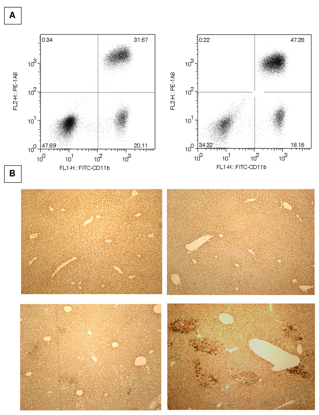
Figure 4. Increased accumulation of neutrophils in the cholestatic livers of NKT cell-deficient mice.
A. The hepatic leukocyte populations were obtained at 18 hours post-BDL and the percentages of Ly-6G+CD11b+ neutrophils (upper right quadrant) constituting the population derived from wild-type (left panels) and Jα18−/− (right panels) mice were determined by flow cytometric analyses. B. The livers of wild-type (left) and iNKT cell-deficient (right) mice were dissected on day 3 following sham-operation (top) or bile duct ligation (bottom). Fixed and paraffin-embedded tissue samples were sectioned and the presence of neutrophils (distinguished by brown precipitate) was assessed by immunohistochemical staining (original 100x magnification). Two experiments yielded comparable results.

