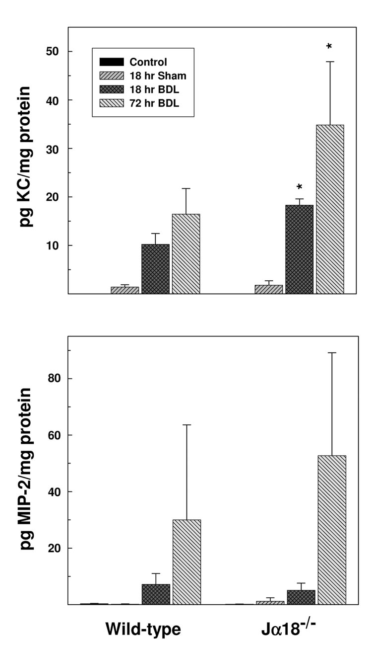
Figure 6. iNKT cells suppress chemokine protein production in the livers of BDL mice.
Representative liver samples were obtained from groups of wild-type and Jα18−/− mice at 0 time (non-operated control), 18 or 72 hours following BDL and/or sham operation. KC (top panel) and MIP-2 (bottom panel) concentrations were determined by Bioplex bead array analysis. Data are the means ± SE derived from 6 mice treated identically. A second experiment yielded comparable results. *Significantly greater than bile duct ligated wild-type mice; P<0.05.

